Structure of Hydra
Hydra has different parts and they function distinctively.
To study the parts very well, let’s look into the structure of hydra, reproductive structure, locomotor structure, and others.
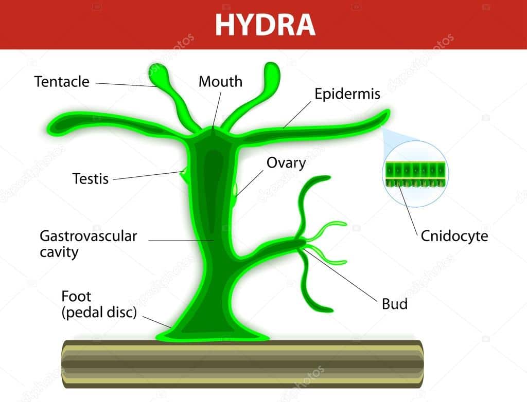
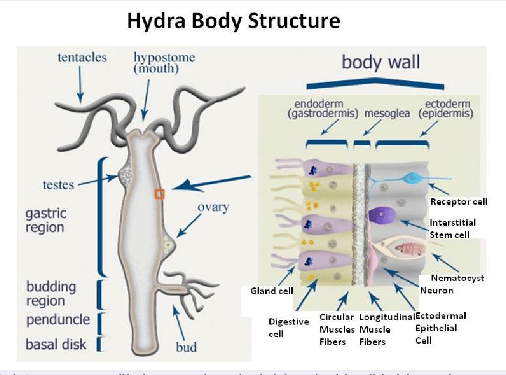
The body of Hydra is cylindrical and is commonly attached to the substratum, for example, rocks or water plants, by the basal closed end which is also known as the foot or basal disc.
The outer end of the body terminates in a small cone-shaped structure called the hypostome, at the apex of which is an opening, the mouth.
This opening leads into a large cavity, the entering which occupies the major portion of the base of the hypostome is a circle of long, hollow tentacles, the cavities of which are continuous with the enteron.
Sometimes two or three rounded knobs may be seen projecting from the surface of the body.
These are the reproductive organ, the ovary, which is situated near the foot while the male reproductive organ, the testis is situated close to the region from which the tentacles grow.
While ovaries occur singly, there may be more than one test at any one time.
Occasionally, a small bud resembling an adult closely in shape is seen growing out from the body near the basal region.
The body wall of a hydra consists of two layers of cells, an outer layer, the ectoderm, and a thicker inner layer, the endoderm.
The ectoderm and endoderm are separated by the mesogloea.
The Ectoderm
The ectoderm is mainly protective and sensory in function. It consists of:
- Musculo-Epithelial Cells.
These form the bulk of the endoderm. they are somewhat columnar with broad outer ends that link up with one another to form a continuous covering for the body.
The cells taper somewhat such that interstices are left in the deeper parts of the ectoderm.
Each cell has a large nucleus. Its inner end is drawn out to form one or more narrow protoplasmic processes that are embedded in the mesoglea and lie parallel to the body surface.
These protoplasmic processes, which are called muscle tails, affect the shape and length of the body.
When they contract, the body becomes shorter and fatter.
- Gland Cells
Certain musculo-epithelial cells in the ectoderm of the foot are also called gland cells because they secrete a sticky substance that enables the foot to attach itself firmly to the substratum.
Other gland cells are found in the hypostome region.
The intake of large pieces of food.
- Interstitial cells
Lying in the interstices between the musculo-epithelial cells are groups of interstitial cells. These are small irregular cells with relatively large nuclei.
They can give rise to any ectodermal cells which have either been damaged or used up.
The interstitial cells also give rise to the reproductive organs and buds by increasing in number very rapidly.
- Cnidoblasts
Cnidoblasts or nematoblasts are very specialized ectodermal cells in each of which lie a nucleus and a large sac, the nematocyst, containing liquid.
The outer end of the sac extends inwards to form a long filament, the nematocyst thread, which is usually coiled into a spiral inside the sac.
A small pointed process, the cnidocil, or trigger hair, projects from the outer side of the cnidoblast.
It is a highly sensitive structure. When it is stimulated by a passing aquatic organism, the protoplasm of the cnidoblast contracts so as to squeeze the nematocysts and cause the coiled nematocyst thread to be forcibly turned inside-out and shot out from the sac.
Cnidoblasts serve mainly for offense and defense, and also for locomotion. There are three kinds of cnidoblasts.
The largest ones have barbs arranged around the bases of the filaments.
When such a cnidoblast is discharged, the barbs wound the victim and the filament penetrates its body, ejecting a poisonous fluid that paralyzes it.
The second type of cnidoblast has no barbs but its filament is coiled spirally.
When the filament is discharged, it coils around the bristles of the prey and helps to hold it securely.
The third type of cnidoblast has a straight, barbless, sticky filament.
The filament adheres to the body of the prey, thus preventing its escape.
Cnidoblasts occur all along the length of the body but especially on the tentacles where they occur in groups called batteries.
Each battery is made up of two or three cnidoblasts of the first type mentioned above, surrounded by several of the other two types.
As the cnidoblasts are continually being used up, they are replaced by new ones which are developed from the interstitial cells.
- Nerve Cells
The never cells are derived from the ectoderm but later penetrate the mesoglea.
Each nerve cell consists of a cell body containing cytoplasm and a nucleus, and several fine, branched processes called nerve fibers.
The ends of the processes meet those of other nerve cells, thus forming a continuous nervous network on the ectodermal as well as the endodermal side of the mesoglea.
The nerve fibers form direct connections with the muscle tails of the musculo-epithelial cells.
- Sensory Cells
Sensory cells are sensitive to touch and it is believed that they also respond to light and chemical stimuli.
They are few and are scattered here and there between the musculo-epithelial cells.
Each cell is narrow and consists of granular cytoplasm and a small nucleus.
At its outer free end is a small, sensitive rod-like projection, while the inner end is prolonged into several branched never fibers which form a synapse with the fibers from the nervous network in the mesogloea.
The Endoderm
The endoderm of Hydra is a less complex but larger layer when compared with the ectoderm.
It serves mainly for direction. It consists of our main types of cells, namely:
- Musculo-Epithelial Digestive Cells
These cells make up the principal part of the endoderm. they are large columnar cells with large nuclei.
Their bases rest on the mesogloea and their other ends project into the enteron.
They function actively in both the shape and size of the body.
At the bases of these cells are muscle tails that are contractile and which run transversely inside the mesogloea.
When these muscle tails contract, the body of the animal becomes longer and narrower.
The inner free ends of some of these cells bear one or two flagella or pseudopodia because of the presence of amoeboids.
The flagella help to maintain the flow of fluid in the enteron while the pseudopodia engulf food particles which are then contained in food vacuoles.
- Gland Cells
The gland cells contain dense cytoplasm and large nuclei and are almost as large as the digestive cells.
However, their bases do not rest on the mesogloea and they do not have muscle tails.
They are most numerous in the hypostome region and the upper part of the body and are responsible for secreting enzymes into the enteron. These enzymes digest food present there.
- Interstitial Cells
Groups of a few small interstitial cells are found at the bases of the larger digestive cells.
The interstitial cells of the ectoderm resemble those of the ectoderm.
- Sensory Cells
Sensory cells similar to those of the ectoderm are scattered in the endoderm.
The Mesoglea
The mesoglea is a thin layer of gelatinous, structureless material.
It is secreted by the ectodermal and endodermal layers and may penetrate between the cells of these layers.
It usually contains nerve cells while the nerve net ramifies over its outer ad inner surfaces.


